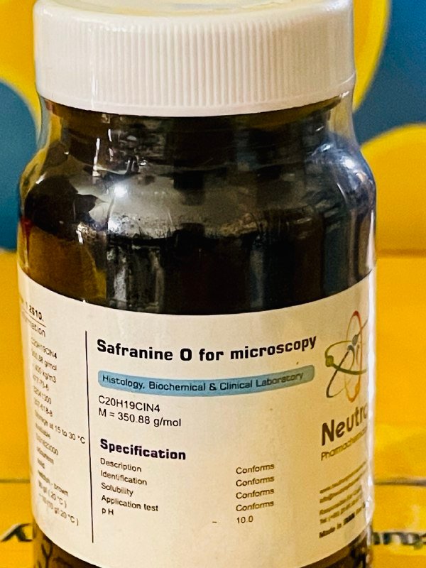
سافرانین او
| Formula | C20H19ClN4 |
| Molar mass | 350.88 g/mol |
| Bulk density | ~ 400 kg/m3 |
| CAS number | 477-73-6 |
| HS Code | 32041300 |
| EC number | 207-518-8 |
| Storage | Storage at 15 to 30 °C |
| SDS | available |
| RTECS | SG1623000 |
| Odour | odourless |
| Form | solid |
| Color | reddish – brown |
| Solubility in water | 50 g/l ( 20 °C ) |
| p H value | ~ 10 (10 g/l 20 °C ) |
| Description | Conforms | ||
| Identification | Conforms | ||
| Solubility | Conforms | ||
| Application test | Conforms | ||
| p H | ~ | 10 |
Safranine O, also known simply as safranin, is a biological stain used widely in histology, cytology, and microbiology. It is a basic dye that binds strongly to acidic (negatively charged) cellular components and is best known for its red or pink coloration of tissues.
🏭⚗️ Production
Safranine O is synthesized as a synthetic basic dye, typically from aniline derivatives through multi-step chemical processes involving diazotization and coupling reactions. It is supplied as a powder or solution, and is soluble in water and ethanol. Stock solutions are usually prepared in distilled water or alcohol, depending on the protocol.
🔬 Properties
Safranine O appears as a bright red powder and yields a pink to red coloration in staining procedures. It binds selectively to acidic tissue components such as nucleic acids and lignified plant cell walls. It has a strong affinity for nuclei and cartilage and functions well as a counterstain due to its contrast with other primary stains. It is pH-sensitive and works optimally in slightly acidic to neutral environments.
🧪 Applications
• Histology: Used as a nuclear counterstain in Gram staining, H&E alternatives, and in various plant tissue protocols.
• Microbiology: Serves as the red counterstain in Gram staining to differentiate Gram-negative bacteria (which stain pink) from Gram-positive ones.
• Botany: Widely used to stain plant tissues—particularly xylem, lignin, and cell walls—in combination with fast green or other contrasting dyes.
• Cytology: Helps visualize nuclei and certain cytoplasmic components in smears and sectioned tissue samples.
• Teaching & Demonstration: Commonly used in educational labs to highlight basic cellular structures for microscope observation.