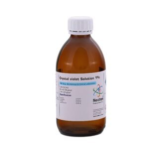
Hematoxyline stain
| Formula | C16H14O6 |
| Density | 1.07 g/cm3 |
| HS Code | 32030010 |
| Storage | at 15 to 25 °C |
| SDS | available |
| Odour | characteristic odour |
| Form | liquid |
| Color | red – violet |
| Solubility in water | soluble( 20 °C) |
| Description | Conforms | ||
| Identification | Conforms | ||
| Solubility | Conforms | ||
| Application test | Conforms |
Hematoxylin stain is one of the most widely used biological dyes in histology and pathology. It is a natural compound derived from the logwood tree and is used to stain cell nuclei blue to purple, typically in combination with eosin in the classic H&E staining method.
🏭⚗️ Production
Hematoxylin itself is not a dye until it is oxidized to hematein, which is then combined with a metal mordant—commonly aluminum, iron, or tungsten salts—to form a staining complex. The resulting solution is typically prepared in water or alcohol, with stabilizers added to extend shelf life. It requires careful preparation and aging (oxidation) to become fully active.
🔬 Properties
Hematoxylin stains nuclei a deep blue or purple due to its binding to nucleic acids in chromatin. It provides excellent contrast for cellular detail, especially nuclear morphology. It is water-soluble, sensitive to pH, and works best when combined with a mordant to bind tissue components effectively. Over time, the stain may degrade or precipitate, requiring filtration or replacement.
🧪 Applications
• Histology: Used to stain cell nuclei in almost all routine tissue sections, typically as the nuclear component of hematoxylin and eosin (H&E) staining.
• Cytology: Highlights nuclear structures in smear preparations, aiding in the evaluation of cell shape, size, and chromatin pattern.
• Pathology: Essential for diagnosis of disease through tissue examination, particularly cancers and inflammatory conditions.
• Special staining protocols: Can be used in regressive or progressive staining techniques depending on the level of contrast desired.
• Teaching & microscopy: Valuable in educational settings for demonstrating cell structure and nuclear organization.


